Archives
-
July
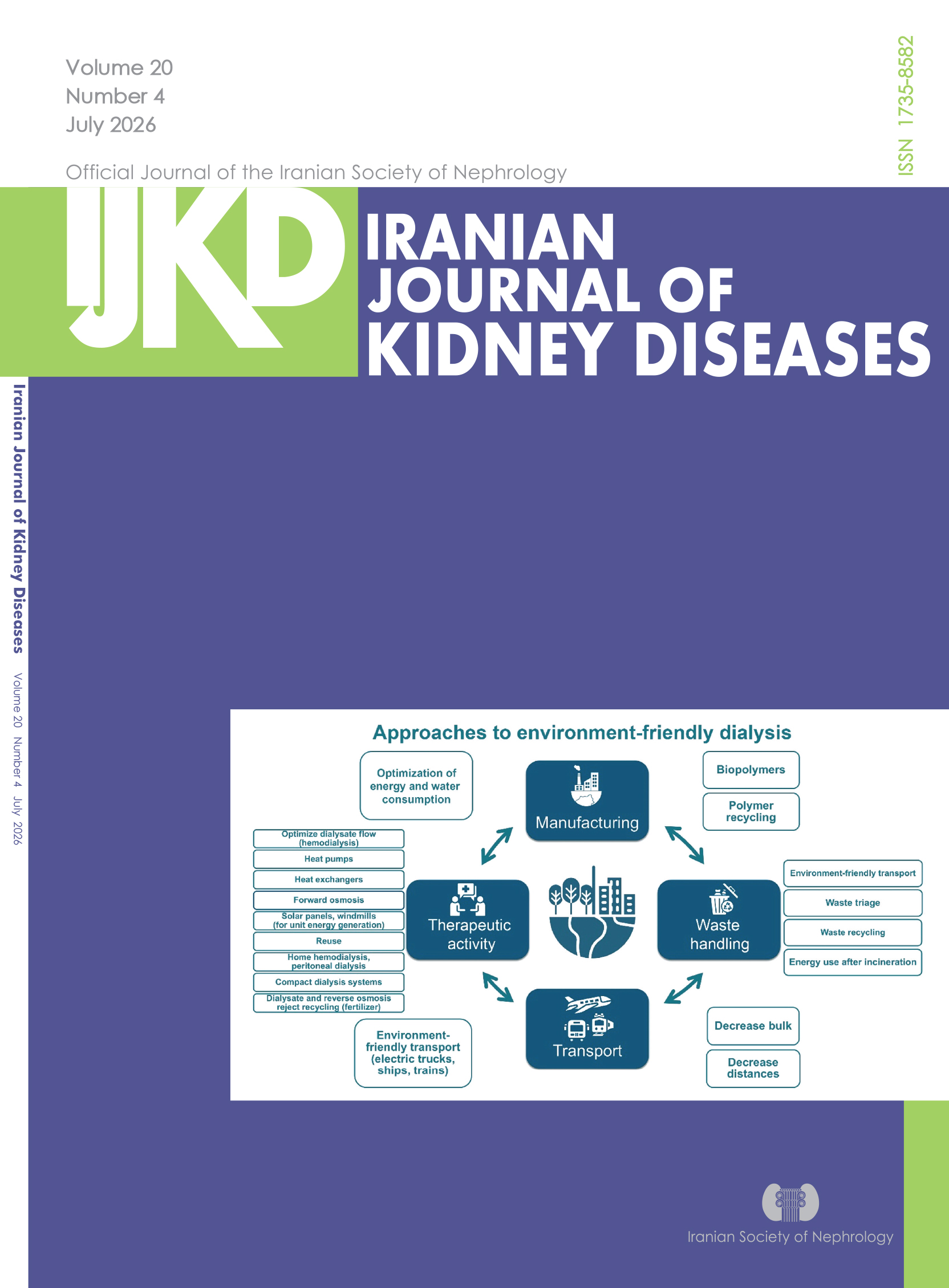
Vol. 20 No. 4 (2026)About the cover: Approaches toward environment-friendly dialysis, categorized for manufacturing, transport, therapeutic activity, and waste handling. The list is not exhaustive. For more detailed lists, see the study by Vanholder et al.6 Environment friendliness should not be pursued at the expense of patient quality of life or clinical status. Compact dialysis systems consume less water or recycle dialysate over sorbents.
-
May
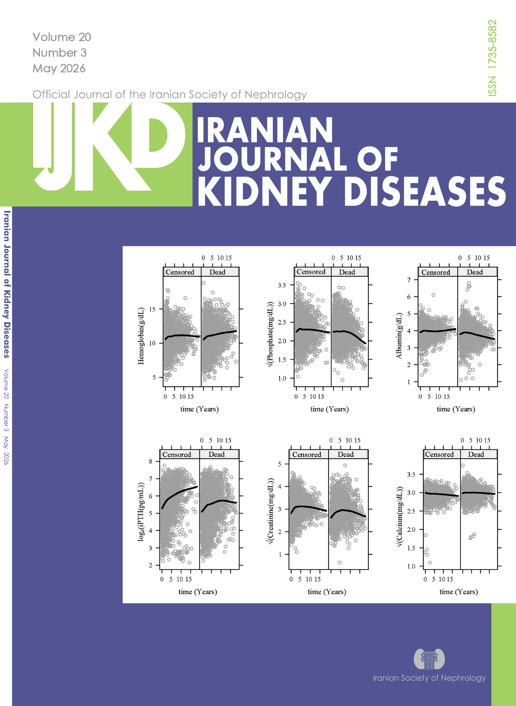
Vol. 20 No. 03 (2026)About the cover:Longitudinal Trajectory Plots Stratified by Survival Status (Censored Vs. Dead). Spline smoothing was used to estimate average trajectories.
-
March
Vol. 20 No. 02 (2026)About the cover:
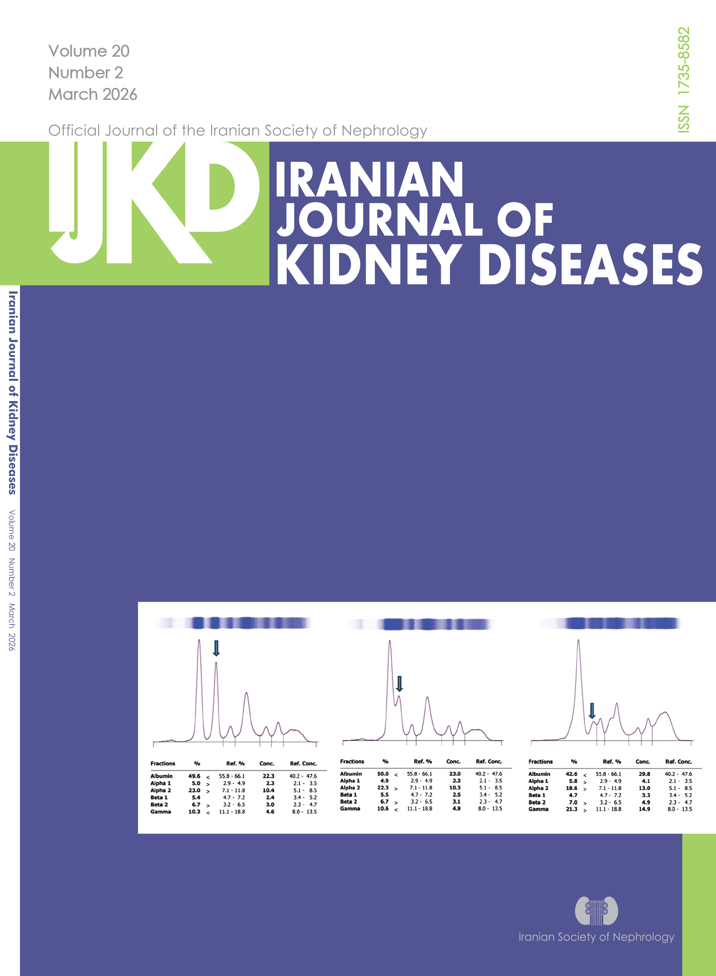
Capillary Serum Protein Electrophoresis (SPEP) Patterns Demonstrating Albumin Fraction Irregularities (AFI) Observed Nephrotic Syndrome Cases
-
January
Vol. 20 No. 01 (2026)About the cover:
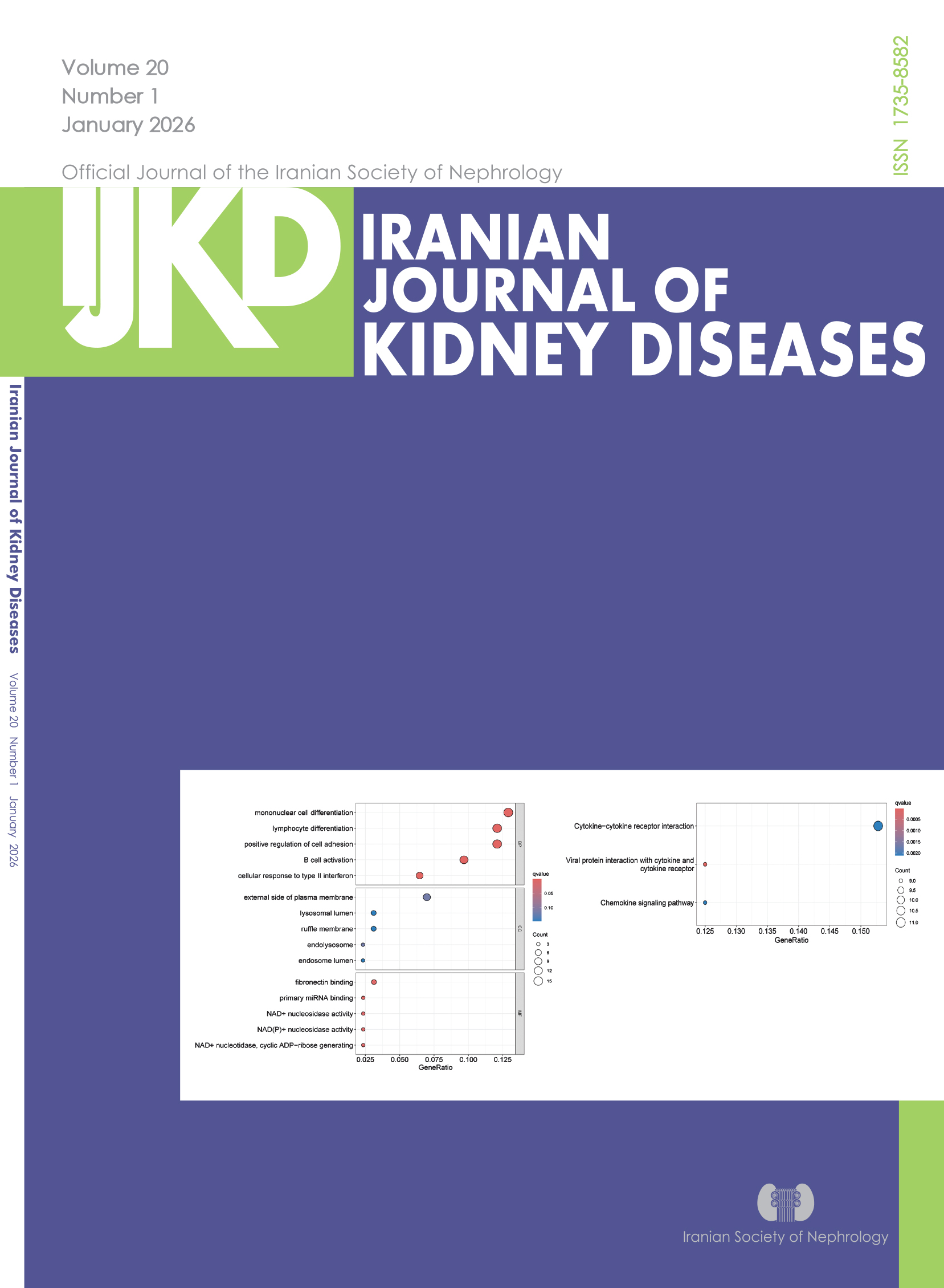
FUMA and Enrichment Analysis of IBD on CKD. Bubble Chart for Gene Ontology (GO) Analysis (Left). Bubble Chart for Kyoto Encyclopedia of Genes and Genomes (KEGG) Analysis (Right).
-
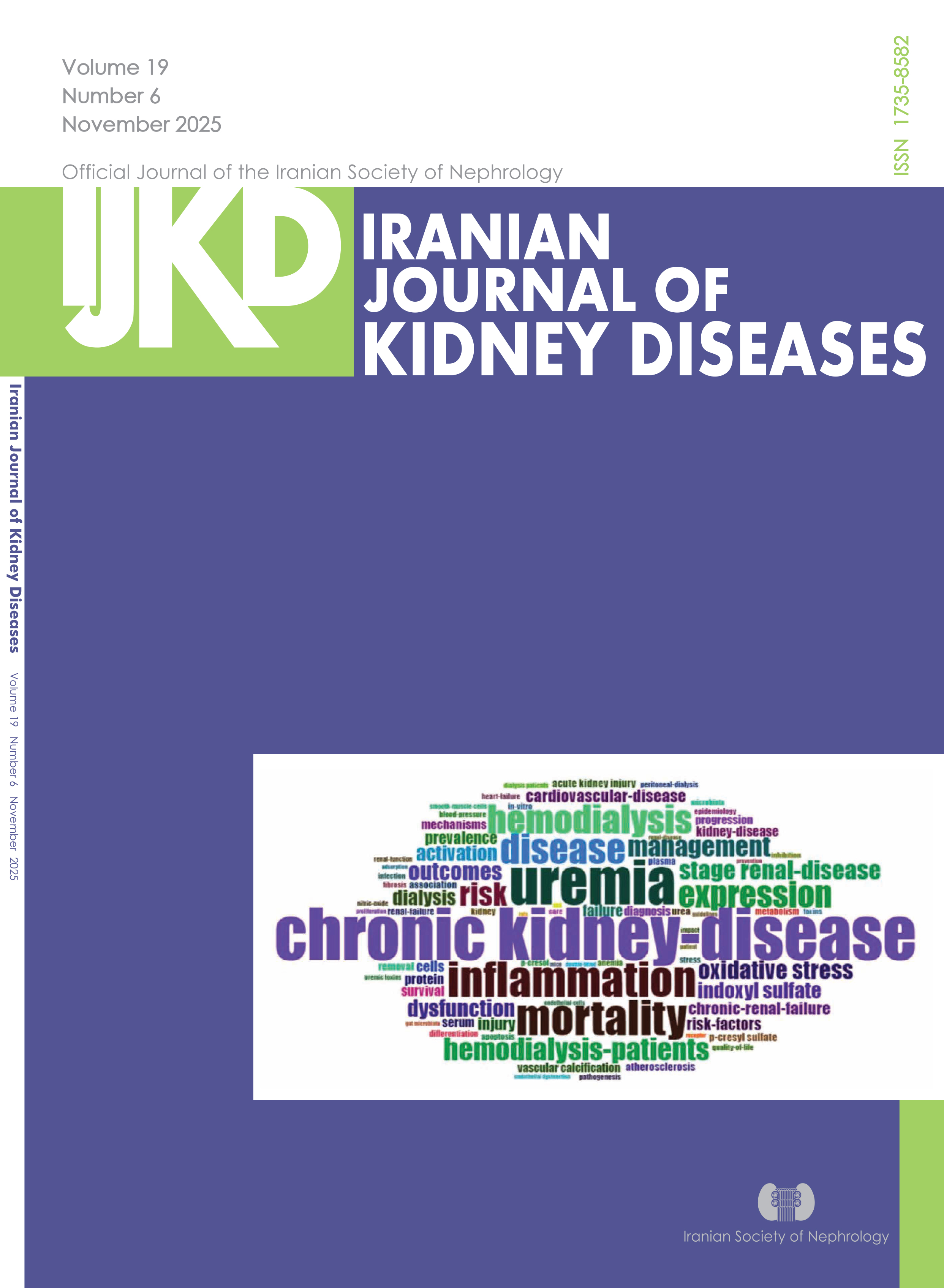
November
Vol. 19 No. 06 (2025)About the cover:
Word Cloud Map Showing the Relative Frequency of Keywords, Where Larger Font Size Corresponds to Higher Frequency.
-
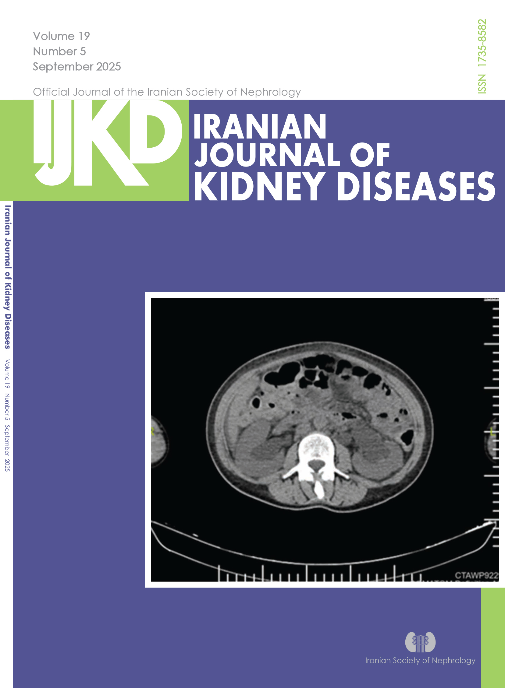
September
Vol. 19 No. 05 (2025) -

July
Vol. 19 No. 04 (2025)About the cover:
Distribution of Responders - white ellipses
Frequency of hyperoxaluria in Iran adopted from literature- Orange Rectangles -

March
Vol. 19 No. 02 (2025)About the Cover:
Astragalus Membranaceus-Salvia Miltiorrhiza Core Target Network
-
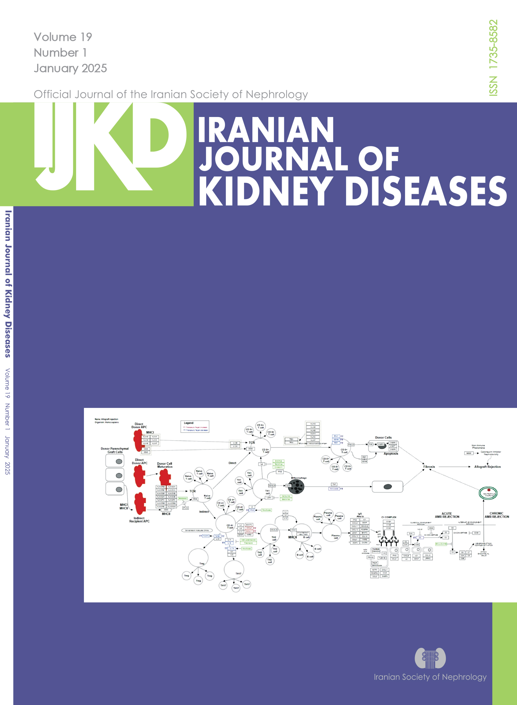
January
Vol. 19 No. 01 (2025) -
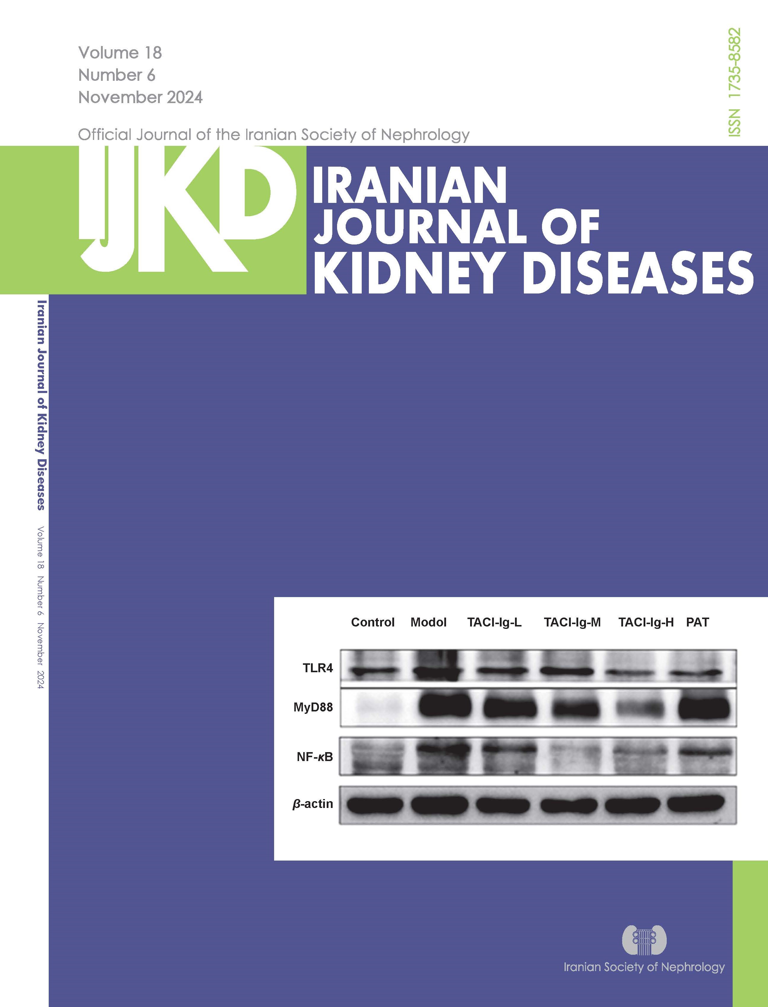
November
Vol. 18 No. 06 (2024)About the cover:
Results of Protein Expresion of TLR4/MyD88/NF-κB; original gel electrophoresis
-
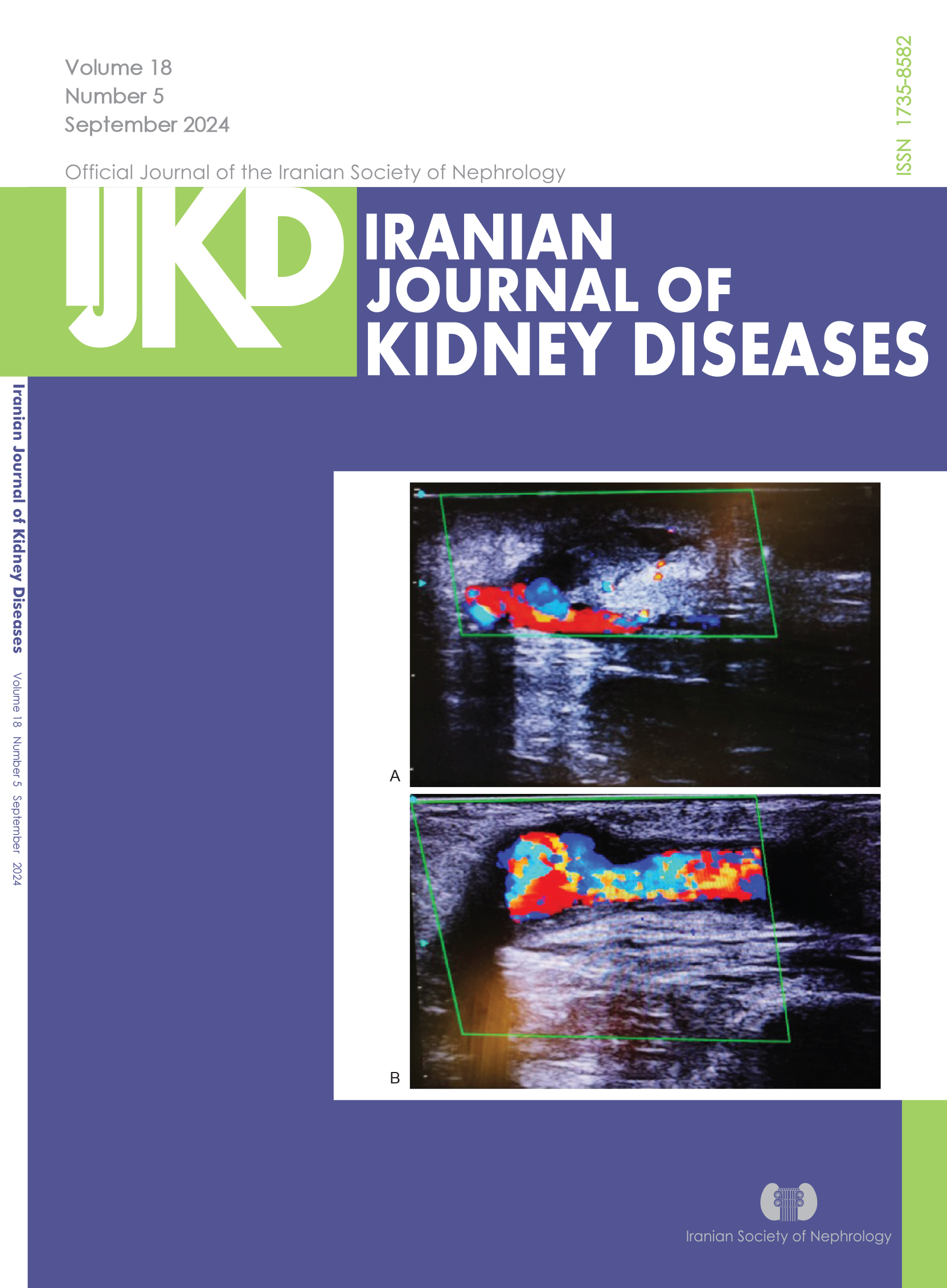
September
Vol. 18 No. 05 (2024)About the cover:
A- Ultrasound Images of Post-thrombolysis AVF
B- Post-PTA AVF, after Thrombolysis
About the cover:
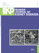
A: SEM images of the interaction between GO (blue arrows), MSCs (green arrows), and macromolecules (yellow arrows)derived from stem cells (Magnification: 2000×), B: green arrows indicate the GO and yellow arrows show macromolecules secreted from
stem cells (Magnification: 2500×) -
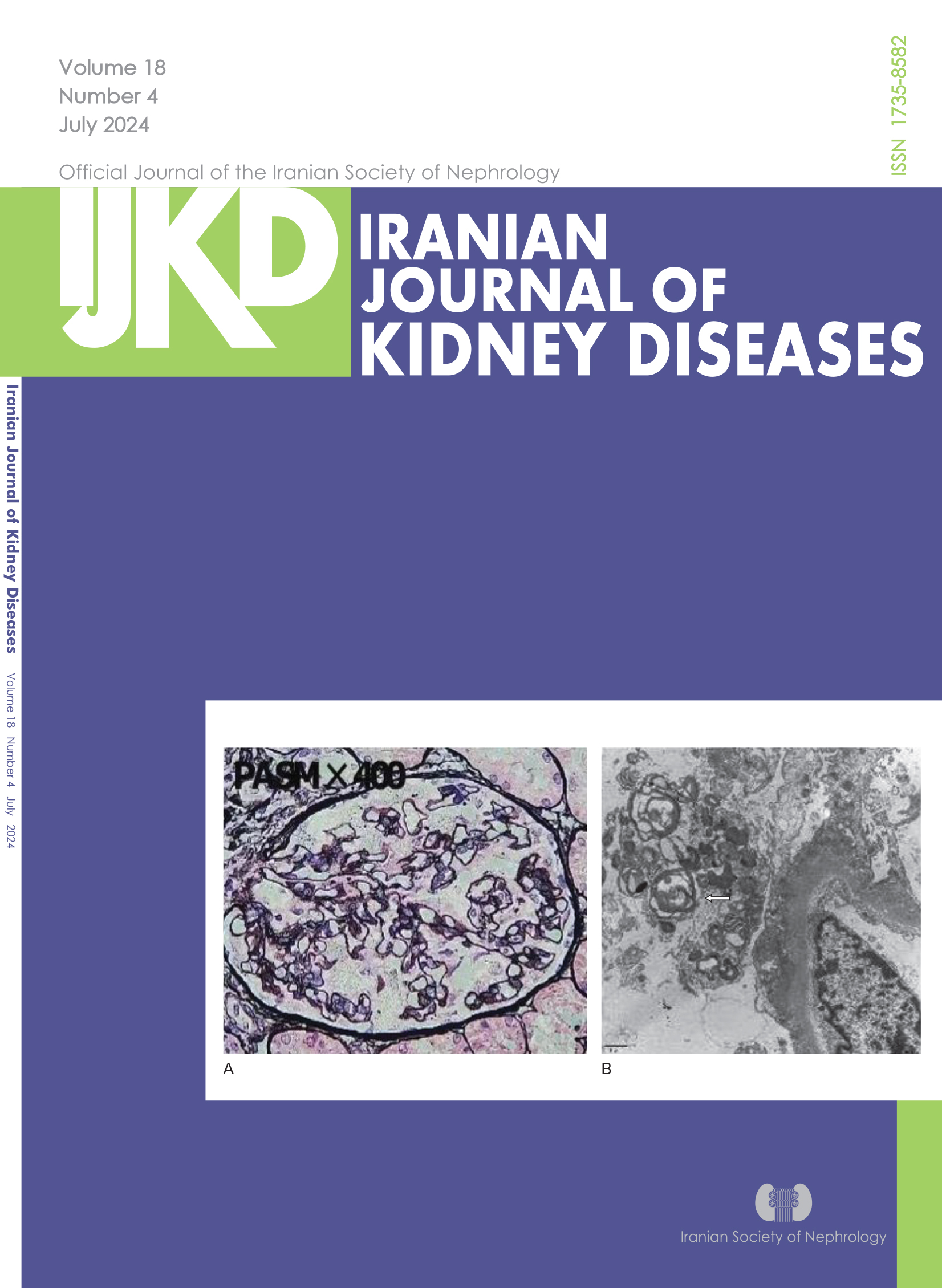
July
Vol. 18 No. 04 (2024)About the cover:
A. Basement Membrane Thickening and Significant Vascular Degeneration of Podocytes.
B. Zebra Bodies in Secondary Lysosomes.
-
May
Vol. 18 No. 03 (2024) -
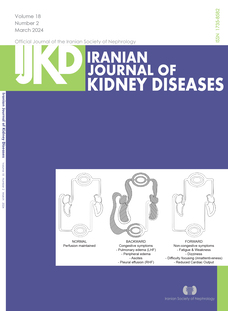
March
Vol. 18 No. 2 (2024)About the Cover:
Comparison of Normal, Congestive, and Hypoperfusive Circulations
-
January
Vol. 18 No. 1 (2024) -
November
Vol. 17 No. 6 (2023)About the Cover
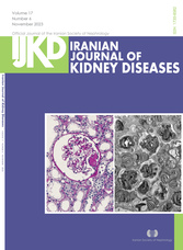
The needle-biopsy specimen of the kidney showed 11 glomeruli(A) , enlarged podocytes contain significant numbers of inclusion bodies that reputed myelin figures and zebra bodies(B).
-
July
Vol. 17 No. 4 (2023)About the cover:
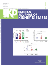
Individual values and mean of (A): BUN level, (B): BUN level differences between group at end of study, and (C): BUN level changes in Hyd. group (blue color), Hyd. + Ator group (red color), Hyd. + Nico (green color) group pre-intervention and after 48 hours angiography-intervention (Abbreviations: BUN, blood urea nitrogen; Hyd, hydration; Ator, atorvastatin; Nico, nicorandil; pre-int., preintervention; After-int., after-intervention)
-
May
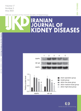
Vol. 17 No. 3 (2023)About the cover: Changes of MAPK and NF-κB Protein Levels in Each Group (Note: 1) sham operation group; 2) Model group; 3) Allicin lowdose group; 4) Allicin medium-dose group; 5) Allicin high-dose group; Compared to the sham group, *P < .05; Compared with the model
group, # P < .05) -
March
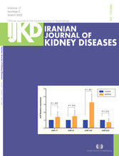
Vol. 17 No. 2 (2023)About the cover: Relative Expressions of the Studied miRs in Control and ADPKD Groups [The ADPKD patients demonstrated significant increases in urinary expressions of miR-17, miR-21, and miR-143. On the other hand, the urine samples of ADPKD patients containedlower levels of miR-223.
-
January
Vol. 17 No. 1 (2023) -
November
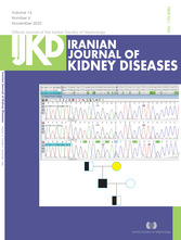
Vol. 16 No. 6 (2022)About the cover:Patient’s deceased mother could be homozygous or heterozygous for NM, and M_144585: exon3:e.568G > C: P.A190P. Chr11:64360938:G > C: gene was confirmed in the patient’s father and son, but NM144585:exon8:e.1301G > C:P.R4341H Chr11:64367854:G > C: variant was only confirmed in
the patient. -
September
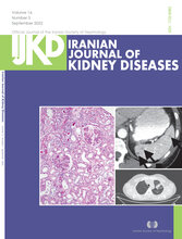
Vol. 16 No. 5 (2022)About the cover: Peribronchial thickening in the left upper lobe of the lung/ Hypodense changes in the spleen
-
July
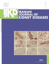
Vol. 16 No. 4 (2022)About the cover:AQP-1, AQP-3, and VEGF-A Staining on Human Peritoneal Mesothelial Cells in the Control, CKD 5, and PD-UFF Groups
-
May
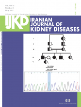
Vol. 16 No. 3 (2022)About the cover: Pedigree of the Family (The black arrow shows the patient with ESKD. p.C113Y mutation in CLDN16 gene with homozygote condition was found in IV-1, IV-3, and IV-4). I-1, II-1, II-2, III-1, III-2, IV-5, and V-1 are obligate carriers.
-
March
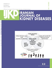
Vol. 16 No. 2 (2022)About the cover: Schematic representation of consumer and health care professionals’ collaborative advocacy using social media platformswith the goal of Kidney Health for All.
-
January
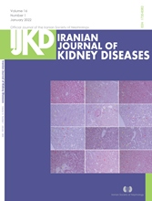
Vol. 16 No. 1 (2022)About the cover: Representative Masson’s trichrome staining (magnification ×400) (A), and H&E staining (magnification ×100) (B) of kidney sections from studied groups received saline, A779, or Losartan. Quantitative (mean ± SEM) analysis of the glomerular injury (C) and inflammtion (D). Red arrows :Glomeruli; yellow arrows: Foci of chronic inflammatory cells aggragation (n = 7 in each group; *** P < .001 vs. sham+ Veh, ## P < .01, ### P < .001 vs. UUO+ Veh, $$ P < .01 vs. UUO+E2)
-
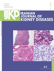
September
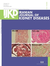
Vol. 15 No. 5 (2021)About the cover: (a)H & E 10×. Glomeruli showing cellular crescents with focal fibrinoid necrosis. Mild acute tubular injury is also noted in the background. (b) JMS 40×. Glomerulus showing cellular crescent formation, double contouring of basement membranes,and focal endocapillary proliferation. (c) PAS 20×. PAS stain showing cellular circumferential crescents with breech of glomerular basement membrane and focal fibrinoid necrosis. Mild acute tubular injury is also noted in the background. (d). Peripheral Blood smear showing schistocytes.
-
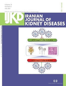
July
Vol. 15 No. 4 (2021)َAbout the cover: Potential effects of SARS-CoV-2 infection on ACE2-Ang-(1-7)-Mas receptor axis in renal tissue
-
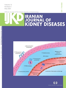
May
Vol. 15 No. 3 (2021)About the cover:
Alterations in the Immune System of Maintained Hemodialysis Patients.Uremic toxins and complement activation are two main causes of the sustained inflammation in hemodialysis patients. Uremic toxins increase the expression and activity of TLR2 and TLR4 in neutrophils and epithelial cells that lead to an increase the in pro-inflammatory cytokine levels. On the other hand, complement-derived anaphylatoxins trigger the release of pro-inflammatory cytokines and cause the activation of endothelium. Sustained inflammation seems to decrease the potential of the adaptive immune system in establishing strong immune responses (Abbreviations: PMNs, polymorphonuclears; AGE, advanced glycation end products).
-
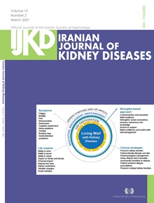
March
Vol. 15 No. 2 (2021)About the cover: Conceptual framework of “Living Well with Kidney Disease” based on patient centeredness and empowering patient with focus on effective symptom management and life participation
-
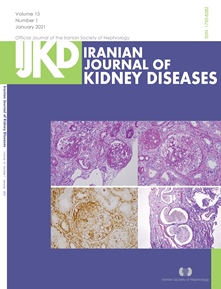
January
Vol. 15 No. 1 (2021)About the cover: A) focal segmental mesengial matrix increase and segemental hyalinosis in two glomerules, Periodic acid shift (PAS)×200, B) glomerular capillary tuft collaps and podocytes proliferation, global sclerosis in one glomerule, Periodic acid shift (PAS);×200, C) staining in proliferated podocytes with Vimentin. IHK;×400, and D) cystic dilatation, tubular atrophy and interstitial fibrosis in tubules. Masson-trichrome; ×200.
-
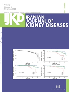
November
Vol. 14 No. 6 (2020) -
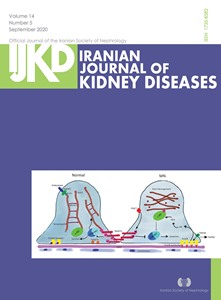
September
Vol. 14 No. 5 (2020)About the cover : Pathways Involved in Podocyte Injury in MN (AA: Arachidonic Acid)
-
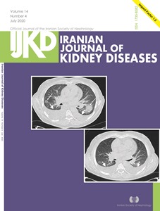
July
Vol. 14 No. 4 (2020)About the cover:A) A 55-year-old male patient with a history of underlying chronic kidney disease (GFR = 9.1, stage V) presented with dry cough and dyspnea which had started since 5 days ago. Initial computed tomography (CT) imaging showed bilateral moderate pleural effusion predominantly in right side, ground glass opacity and area of consolidation in middle zones. B) CT imaging obtained 22 days later show bilateral mild pleural effusion and complete lesion absorption. The patient was discharged after 7 days.
-
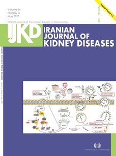
May
Vol. 14 No. 3 (2020)About the cover: Schematic representation of viral entry to the host cell, processing within the cell and virus-induced immune response. Some potential drugs and their targets are also presented.
-
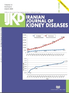
March
Vol. 14 No. 2 (2020)About the cover: a)It demonstrates incidence and prevalence of HD patients in Iran during the past 2 decades. b)It mentions new patients added to maintenance hemodialysis pool.
-
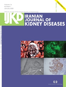
January
Vol. 14 No. 1 (2020)About the cover: a)This figure shows Jones’ staining of a paitient with membranous nephropathy (×40). b) total Ig. c)It determines electron microscope study in a patient with membranous nephropathy. d)Immunohistochemistry with anti-C4d (polyclonal) in MGN reveals positive immunoreactivity along BM with granular pattern.
-
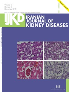
November
Vol. 13 No. 6 (2019)About the cover:Cross-sectional view of the kidneys showing the size of Bowman’s space and necrosis in groups: A, No intervention control; B, Sham operation without vascular obstruction and receiving the PBS; C, 45 min ischemia and 24h reperfusion and receiving PBS; and D, 45 min ischemia and 24h reperfusion and receiving 2× 10⁶ AD-MSC. Magnification x 400, hematoxylin-eosin staining.
-

Supplement 1
Vol. 13 (2019)Abstracts of the 17th International Congress of the Nephrology, Dialysis, and Transplantation that was organized by Iranian Society of Nephrology. -
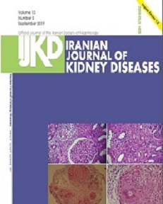
September
Vol. 13 No. 5 (2019) -
July
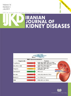
Vol. 13 No. 4 (2019)About the cover:Mesenchymal stem cells have immunomodulatory properties through their paracrine effects with different mechanisms. MSCs predominantly improve renal regeneration by secretion of different growth factors, cytokines and chemokines which decrease inflammation, apoptosis, and fibrosis. MSCs: Mesenchymal stem cells; DCs: Dendritic cells; MO: Monocytes; MΦ:Macrophages; Treg cells: Regulatory T cells; Th: T helper; Nk cells: Natural killer cells; PMN: polymorphonuclear neutrophils PGE2: prostaglandin E2; IDO: indoleamine 2, 3-dioxgenase; TGFβ: transforming growth factor; HGF: hepatocyte growth factor; IL: Interleukin; TNFα: tumor necrosis factor-α; iNOS: inducible nitric-oxide synthase; HO1: haem oxygenase-1; sHLA-G5: soluble HLA-G5; NO: nitric oxide; TSG-6: TNF-stimulated gene 6 protein; IFN-γ: interferon gamma.
-
May
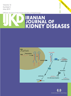
Vol. 13 No. 3 (2019)About the cover: The role of miR-200 family down regulation by Dicer inactivation on PKD development. The dicer inactivation caused down regulation of kidney tissue specific miRNAs and thus activation of PKD1.
-
March
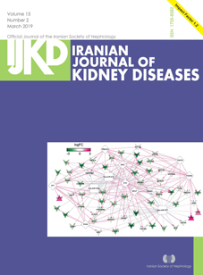
Vol. 13 No. 2 (2019)About the cover: Network of co-expressed differentially expressed genes (DEGs) with consistency. The green lines represent the connection of negative correlation gene pairs, while the red lines represent the connection of positive correlation gene pairs. The change of nodes color from green to red presents the change of logFC from negative to positive. The regular triangle and inverted triangle present significantly up-regulated DEGs and down-regulated DEGs respectively.
-
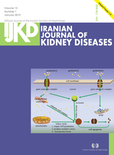
January
Vol. 13 No. 1 (2019)About the cover:The underlying mechanics of antibiotic-induced acute kidney injury. Antibiotics are a wide-spread class of drugs and the principal cause of antibiotics -associated nephropathy varied. Different mechanisms have been identified: a decrease in kidney perfusion pressure, the formation of encounter complex, cytotoxicity, hypersensitivity reactions and crystalline nephropathy. ATN indicates acute tubular necrosis; AIN, acute interstitial nephritis; and AKI, acute kidney injury.
-
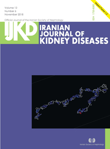
November
Vol. 12 No. 6 (2018)About the cover:Predicted 3D structure model of mutant protein encoded by c.1204C > T (p. H402Y) variation of complement factor H.
-
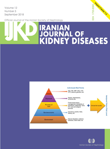
September
Vol. 12 No. 5 (2018)About the cover:Acute kidney injury risk stratification. Adapted from the Acute Disease Quality Initiative with permission.3 DM indicates diabetes mellitus; HBP, high blood pressure; CKD, chronic kidney disease; CHF, congestive heart failure; CLD, chronic liver disease; and ICU, intensive care unit.
-
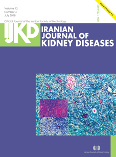
July
Vol. 12 No. 4 (2018)About the cover:The histopathological and immunohistochemical findings of the renal specimen. Left, Fibrotic lesions and inflammatory cells infiltration in the interstitial compartment (hematoxylin-eosin, × 200). Middle, Storiform fibrosis in the interstitial compartment (periodic acid-silver metheramine, × 200).
-
May
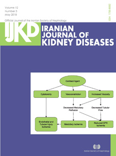
Vol. 12 No. 3 (2018)About the cover:Different mechanisms through which contrast agents may induce contrast nephropathy
-
March
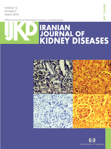
Vol. 12 No. 2 (2018)About the cover:Rats treated with dexamethasone, vitamin E, and P major extract (600 mg/kg and 1200 mg/kg) showed mild tubular, glomerular, and interstitial changes (hematoxylin-eosin, × 400).
-
January
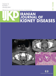
Vol. 12 No. 1 (2018)About the cover: A 26-year-old man, known case of urinary calculus disease, presented with the chief complaint of 1 episode of hematuria
-
November
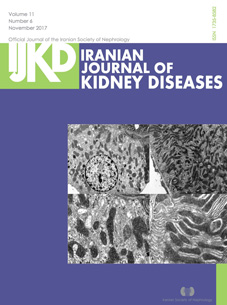
Vol. 11 No. 6 (2017)About the cover:Ultrastructural alteration in renal tubular epithelial cells mitochondria in 5/6 nephrectomized rats
-
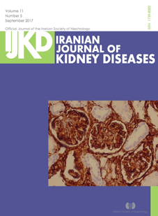
September
Vol. 11 No. 5 (2017)About the cover: Vimentin diffusely stained glomerular, tubular components, and stroma (vimentin, × 400).
-
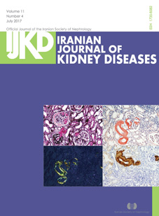
July
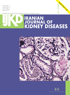
Vol. 11 No. 4 (2017)About the cover:A, Light microscopy showing nodular mesangial expansion and arteriolar thickening with silver-negative materials. B, Congo-red stain showing pink-reddish mesangial and vascular staining. C, A section showing typical apple-green birefringence under polarized light microscopy. D, Immunohistochemical staining for serum amyloid A is positive.
-
Supplement 1
Vol. 11 (2017)Abstracts of the 3rd International Congress of Organ
Transplantation & the16th International Congress of Nephrology, Dialysis, and Transplantation
-
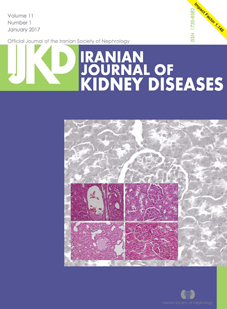
January
Vol. 11 No. 1 (2017)About the cover: Tissue necrosis in the rats with 45 mg/kg of paraquat-Preserved tubules and a glomeruli in the rats with 45 mg/kg of paraquat and ellagic acid-Glomerular necrosis (arrow) in the rats with 15 mg/kg of paraquat-Improved tubular and glomerular histology in the rats with 15 mg/kg of paraquat and ellagic acid.
-
November
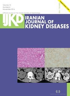
Vol. 10 No. 6 (2016)About the cover: Computed tomography revealed a solid, well-circumscribed lesion in the right kidney. Left, The mass was not delineated on a pre-enhanced scan. Right, The mass was moderately enhanced on an enhanced scan-Left, Tumor cells exhibited uniform round nuclei with clear-to-eosinophilic granular cytoplasm (hematoxylin-eosin, × 400). Right, Tumor cells demonstrated diffuse strong positivity to CD34 (immunostaining).
-
September
Vol. 10 No. 5 (2016) -
July
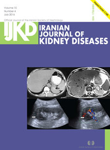
Vol. 10 No. 4 (2016)About the cover:A 41-year-old nondiabetic nonhypertensive man presented with increased frequency of micturition and low-grade fever.
-
May
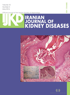
Vol. 10 No. 3 (2016)About the cover: Pathologic examination shows central cup-shaped invagination of the epidermis consisting of keratin plug admixed with parakeratotic basophilic cellular debris. The inflammatory infiltration of granulocytes is observed in the dermis. Orcein staining for elastic fibers was negative.
-
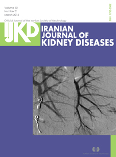
March
Vol. 10 No. 2 (2016)About the cover: Multiple diffused microaneurysm in renal parenchyma using digital subtraction angiography-The feeding artery to the pseudoaneurysm was completely ligated using 3 Tornado microcoils.
-
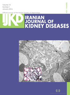
January
Vol. 10 No. 1 (2016)About the cover:A large number of myeloid bodies (zebra bodies, black arrow) were visible via electron microscopy in the glomerular epithelial cytoplasm. The basement membrane appeared slightly but irregularly thickened. Epithelial cells showed diffuse effacement of the foot processes, with few electron-dense deposits visible under the epithelium (× 5000; scale bar, 2 μm).
-
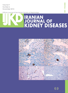
November
Vol. 9 No. 6 (2015)About the cover:Deletion of cyclic AMP response element-binding protein (CREB) in renin cells does not alter basal renin expression in adult kidney, but influence renin recruitment in low-sodium diet mice kidney. Top two rows are immunostaining for renin and CREB in consecutive sections of kidneys from wild type and CREB gene knock out mice, respectively. In wild type kidney, renin cells are positive for CREB (brown nuclei), but in CREB knock out kidney, CREB is absent (blue nuclei). Bottom two rows are immunostaining for renin in wild type and CREB knock out mice with normal and low-sodium diet, respectively. In wild type mice, renin staining increased in low-sodium diet kidney compared with normal diet. By contrast, in CREB knock out mice renin staining did not increase or even decreased in low-sodium diet kidney.
-
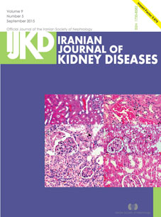
September
Vol. 9 No. 5 (2015)About the cover: Histopathological assessment (hematoxylin-eosin). A, Normal renal tissues of group 1. B, Homogenous hyaline thickening of the renal blood vessels wall (arrows) in group 2 (cyclosporine). C, Cystic dilatation of the Bowman capsule (asterisks), moderate vacuolation of the distal convoluted renal tubules (arrows), and few pyknotic nuclei of renal epithelium (arrowheads) in group 2 (cyclosporine). D, Moderate vacuolation of renal tubules (arrows) and few pyknotic nuclei of renal epithelium (arrowheads) in group 3 (cyclosporine and olive leaf extract, 40 mg/kg). E, Shrunken glomerular tufts (arrows) with mild interstitial nephritis represented by few fibroblasts and mononuclear cell infiltrations (asterisks) in group 4 (cyclosporine and olive leaf extract, 80 mg/kg). F, Mild dilatation of the Bowman capsule with restoration of the normal histological features of the renal tissue in group 5 (cyclosporine and olive leaf extract, 120 mg/kg).
-
July
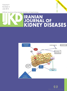
Vol. 9 No. 4 (2015)About the cover:Transmembrane proteins and their activation. CHOP indicates CCAAT/enhancer-binding protein homologous protein; Nrf2, nuclear factor (erythroid-derived 2)-like 2; ATF4, activating transcription factor 4; XBP1, X-box binding protein 1; IRE1, nositol-requiring transmembrane kinase/endonuclease-1; ATF6, activating transcription factor 6; and ERAD, endoplasmic reticulum-associated protein degradation.
-
May
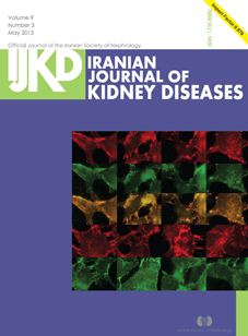
Vol. 9 No. 3 (2015)About the cover:Changes in distribution of β-catenin and P-cadherin in diabetic conditions and vitamin D treatment (× 1000). β-Catenin was colocalized with P-cadherin at cell membrane areas. Top, Diabetic conditions (B30, A5, and A30) decreased the immunostainings and disrupted the distributions of β-catenin and (arrows). Middle and Bottom, Changes of β-catenin and P-cadherin were improved by vitamin D.
-
March
Vol. 9 No. 2 (2015) -
January
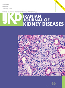
Vol. 9 No. 1 (2015)About thr cover:Pathology of drug-induced acute interstitial nephritis. Light microscopy demonstrating acute interstitial nephritis with inflammatory cells, interstitial edema, and tubulitis. Red arrows identify tubulitis while the white arrows point out eosinophils
(hematoxylin-eosin, × 40). -
November
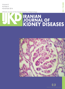
Vol. 8 No. 6 (2014)About the cover: Glomerular capillary tuft segmentation and capillary loop closure with meningeal cell proliferation and leukocytes infiltration without extra capillary proliferation (crescent) formation.
-
September
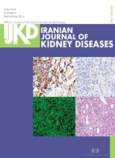
Vol. 8 No. 5 (2014)About the cover:Tubulointerstitial nephritis accompanying γ-heavy chain deposition and γ-heavy chain restricted plasma cells in the kidney.
-
July
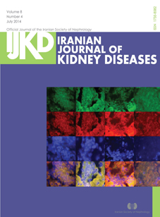
Vol. 8 No. 4 (2014)About the cover: Immunofluorescent staining of α-actinin and β-catenin. Double-staining of α-actinin and β-catenin show a decrease in the foot processes of podocytes around capillary loops in rat glomeruli at 10 weeks after diabetic condition (× 400).
-
May
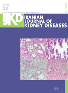
Vol. 8 No. 3 (2014)About the cover: A, Interstitial inflammation with tubular atrophy and calcium deposit (hematoxylin-eosin). B, Extensive interstitial and tubular calcification. C, Trichorom staining shows interstitial fibrosis. D, von Kossa staining.
-
January
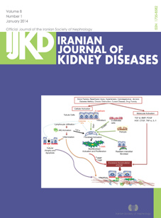
Vol. 8 No. 1 (2014)About the cover:Mechanisms and mediators of renal interstitial fibrosis and tubular atrophy (IFTA).Immunological (acute rejection, previous transplantation, HLA mismatch) and nonimmunological (donor’s age, source, reperfusion injury, hypertension, diabetes, chronic obstruction, bacterial pyelonephritis, viral nephritis, and drug toxicity) factors trigger renal fibrosis. Transforming growth factor-β induces matrix production through Smad3-dependentpathway and inhibits extracellular matrix degradation by suppressing MMPs and inducing the inhibitor of matrix metalloproteinases (tissue inhibitor of metalloproteinases). Moreover, it induces myofibroblast formation through tubular epithelial–mesenchymal transition. Cellular (epithelial-mesenchymal transition, fibroblast activation, lymphocyte infiltration, and cellular apoptosis) and molecular activations lead to the excessive accumulation of ECM. TGF-β indicates transforming growth factor-β; BMP, bone morphogenetic protein; PDGF, Platelet-Derived Growth Factor; HGF, hepatocyte growth factor; CTGF, connective tissue growth factor; TNF-α, tumor necrosis factor alpha-α; IL-1, interleukin 1; EMT, epithelial-mesenchymal transition; EndMT, endothelial-to-mesenchymal transition; and SMA, smooth muscle actin; MMP, matrix metalloproteinases; TIMP, tissue inhibitor of metalloproteinase; and ECM, extracellular matrix.
-
November
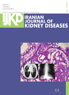
Vol. 7 No. 6 (2013)About the cover: One glomerulus shows a sclerotic segment with adhesion to the Bowman capsule (periodic acid–Schiff, × 400)-Chest computed tomography scan of the patient shows bilateral bronchiectasis-Chest radiography of the patient shows dextrocardia and gastric air in the right subdiaphragm.
-
September
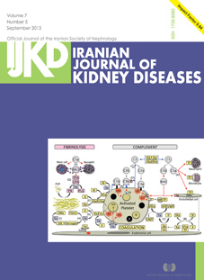
Vol. 7 No. 5 (2013)About the cover:Interplay between complements and the coagulation system. Complement components are labeled by Arabic numerals in grey circles. Coagulation factors are indicated in yellow rectangles except for thrombin, which is in a purple rectangle. A lower case a following Roman numerals stands for activated. An inhibitory effect is indicated by an inverted T sign. A red star indicates an activating effect. See text for details. PAI-1 indicates plasminogen activator inhibitor-1; TF, tissue factor; TPA, tissue plasminogen activator; UPA, urokinase plasminogen activator; FDP, fibrin degradation products; PLG, plasminogen; PN, plasmin; PS, P-selectin; MP, microparticle; VWF, Von Willebrand factor; α, platelet alpha granule; δ, platelet delta granule; and PAR, protease-activated receptor.
-
July
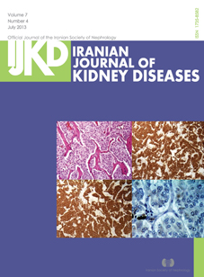
Vol. 7 No. 4 (2013)About the cover:Histopathology of the resected adrenal adenoma. A, Nests of tumor cells sepatated by fibrovascular stroma, a typical Zellballen pattern (hemaoxylin-eosin); B, Positive chromogranin stain; C, Positive synaptophysin stain; and D, negative S-100 protein stain.
-
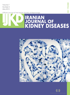
May
Vol. 7 No. 3 (2013)About the cover:Brown-stained Ki-67 positive cells within the glomerular capillary tuft in class II lupus nephritis (Streptavidin-biotin peroxidase staining, × 400).
-
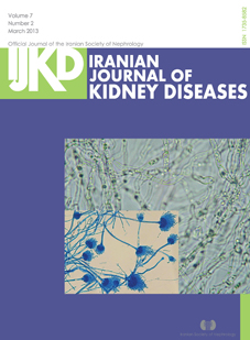
March
Vol. 7 No. 2 (2013)About the cover : A direct examination of the removed fungus ball showing septate hyphae and branching at acute angles, suggestive of Aspergillus (KOH Preparation, × 100)- Biseriate conidial head and conidiophore of Aspergillus terreus (Lactohenol aniline blue preparation).
-
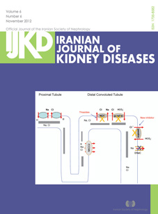
November
Vol. 6 No. 6 (2012)About the cover:The synergistic effect of Na-K-2Cl cotransporter and Na-Cl cotransporter inhibition by furosemide and thiazides, respectively, significantly increases the magnitude of salt excretion. Pendrin expression is increased in response to increased delivery of salt to the collecting duct in order to mitigate the amount of salt wasting. Epithelial sodium channel indicates ENaC; NKCC2, Na-K-2Cl cotransporter; and NCC, Na-Cl cotrasporter.
-
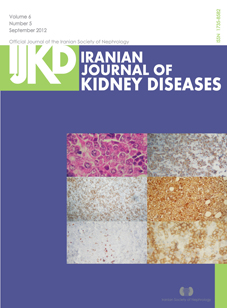
September
Vol. 6 No. 5 (2012)About the cover:Biopsy shows malignant neoplastic tissue composed of large cells with vesicular nuclei, small to indistinct nucleoli, and abundant amphophilic to eosinophilic cytoplasm. Scattered cells resembling immunoblasts with large eosinophilic nuclei were also noticed. Immunohistochemical staining was positive for epithelial membrane antigen (EMA), vimentin, CD79a, CD138, and CD38.
-
July
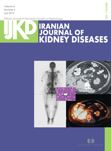
Vol. 6 No. 4 (2012)About the cover: Abnormal soft tissue uptake in the inferior lateral aspect of the right kidney highly suspicious for malignancy- Pre-operative computed tomography of the abdomen showing a 16 × 15-cm heterogeneous multilobulated right renal mass-positron emission scan shows fludeoxyglucose-avid masses in the right nephrectomy bed, anterior abdominal wall and peritoneum highly suggestive of recurrence of the tumor and metastatic disease.
-
May
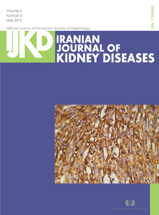
Vol. 6 No. 3 (2012)About the cover: Immunoreactivity for vimentin in neoplastic spindle cells and vascular endothelial lining.
-
March
Vol. 6 No. 2 (2012) -
January
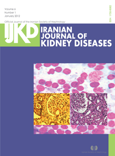
Vol. 6 No. 1 (2012)About the cover:Bone marrow aspiration showed diffuse involvement of marrow with plasma cells-Second kidney biopsy, 1 year after treatment of multiple myeloma showed chronic tubulointerstitial nephritis-First kidney biopsy.
-
Supplement 2
Vol. 5 (2011)Abstract Book of the 13th Congress of Nephrology, Dialysis, and Transplantation -
November
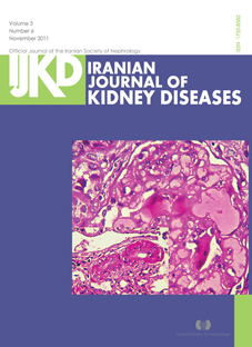
Vol. 5 No. 6 (2011)About the cover:Chronic kidney disease in a child with atypical hemolytic uremic syndrome. Some glomerular capillary lumens contain fibrin thrombi, a few are closed as a result of thickening of capillary walls, and some are ectatic because of mesangiolysis (periodic acid shift, × 400).
-
September
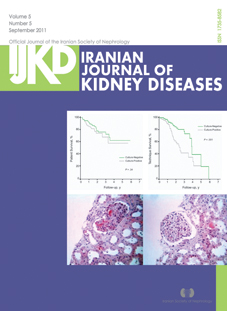
Vol. 5 No. 5 (2011)About the cover: Comparison of patient and technique survival in patients with culture-positive and culture-negative peritonitis.
-
Supplement 1
Vol. 5 (2011)Abstracts of the 2nd International Congress of the Iranian Society of Pediatric Nephrology -
July
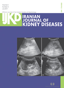
Vol. 5 No. 4 (2011)About the cover:Abdominal ultrasonography. A, A mildly enlarged left kidney without a mass, and hydronephrosis or perinephric fluid collection on day 2. B, An enlarged left kidney with a 4-cm ill-defined hyperechogenic mass with loss of corticomedullary junction of the kidney at the left upper pole on day 5. C, An unchanged left renal mass without the evidence of abscess formation on day 10. D, Complete disappearance of the renal mass with a normal kidney size and echogenicity on day 14.
-
May
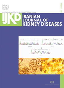
Vol. 5 No. 3 (2011)About the cover: Sequence profiles of CD86 +1057GA genotypes-An example of Wilms tumor with unfavorable histology showing strong positive immunostaining for p53 (anti-p53, × 25 and ×100).
-
March
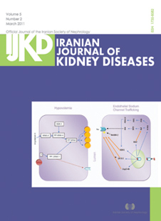
Vol. 5 No. 2 (2011)About the cover:Action of aldosterone in hypovolemia. Elevated level of angiotensin II in the setting of hypovolemia induces WNK-4 phosphorylation, which in turn phosphorylates SPAK (serine threonine proline-alanine rich kinase). Phosphorylated SPAK activates Na/Cl cotransporter. On the other hand, an unknown factor phosphorylates LWNK-1 which downregulates ROMK channel. Therefore, potassium secretion is inhibited despite elevated aldosterone level-Role of aldosterone in endothelial sodium channel (ENac) trafficking. Aldosterone induces serum and glucocorticoid-regulated kinase 1 (SGK1), 14-3-3, Usp2-
45, glucocorticoid-induced leucine zipper and K-ras. K-ras downregulates ENac via Raf pathway. On the other hand, it upregulates ENac by phosphorylating SGK1. Serum and glucocorticoid-regulated kinase 1, ubiquitin-specific protease (Usp2-45), and 14-3-3 deubiquitylate ENac. neural precursor cell-expressed developmentally downregulated (gene 4) protein (Nedd4-2) ubiquitylates ENac. -
January
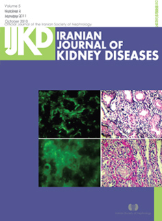
Vol. 5 No. 1 (2011)About the cover: Indirect immunofluorescence with anti-IgG antibody in control specimen showing no positivity (× 200). indirect immunofluorescence with anti-IgG antibody with focal cytoplasmic staining in cortical tubular cells-lymphocytic infiltration of interstitium, making tubulitis (periodic acid-Schiff, × 400). Right, histiocytic aggregation in the interstitium compatible with granuloma formation (Jones, × 400).