Archives - Page 2
-

July
Vol. 4 No. 3 (2010)About the cover: Pathological grading of the rat kidneys after reperfusion injury. A, Grade 1 ischemic injury with brush border loss (periodic acid-Schiff, × 200). B, Grade 2 injury showing brush border loss and tubular dilatation (periodic acid-Schiff, ×100). C, Grade 3 injury with tubular dilatation and necrosis (hematoxylin-eosin, × 100). D, Grade 4 injury with widespread necrosis (periodic acid-Schiff, × 100).
-
April
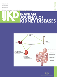
Vol. 4 No. 2 (2010)About the cover: Effect of dietary phosphorus load on phosphorus metabolism in the body.43
1,25(OH)2D indicates 1,25-dihydroxyvitamin D3; Ca2+, calcium; FGF-23, fibroblast growth factor-23; FGFR, fibroblast growth factor receptor; PO42-, phosphate; PTH, parathyroid hormone.-Factors that affect phosphorus absorption7 along the gastrointestinal tract with varying pH.37 -
January
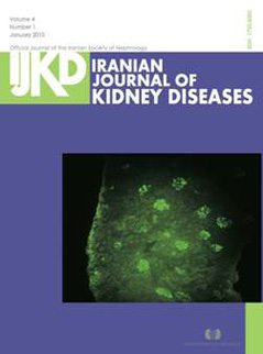
Vol. 4 No. 1 (2010)About the cover:Immunfluorescence microscopy showed mesangial staining for IgA (FITC antihuman IgA, × 400)
-
Supplement
Vol. 3 (2009)Abstract Book of the 12th Congress of Nephrology, Dialysis, and Transplantation -

October
Vol. 3 No. 4 (2009)About the cover:Marked endocapillary hypercellularity with lobular accentuation characterized by mesangial cell proliferation and leukocytes infiltration-Thickening of the glomerular basement membrane with double contour appearance in silver stained sections. Intraluminal plugs are also present-Amorphous, eosinophilic, fuschinophilic positive deposits totally filling the capillary lumina (intraluminal thrombi or cryoplug)-ntense, massive staining of immunoglobulin M with huge deposits filling the capillary lumen.
-

July
Vol. 3 No. 3 (2009)About the cover: Glomeruli show mesangial widening with hypercellularity (hematoxylin-eosin)-Immunofluorescent antibody technique study revealed deposition of immunoglobulin A and complement 3 in the mesangial regions.
-
April
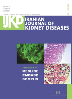
Vol. 3 No. 2 (2009)About the cover: Immunofluorescent antibody technique study revealed a typically full-house pattern-Diffuse prilferative lupus nephritis (stage IV-G) with cellular crescent formation and interstitial infiltration-Proliferative glomerulonephritis with wire loops and intraluminal thrombi (arrows).
-

January
Vol. 3 No. 1 (2009)About the cover: A 20-year-old man presented with elevated serum creatinine level 3 months after kidney transplantation. Cytomegalovirus (CMV) pp65 antigen was detected by indirect immunofluorescent method. Figure A shows contaminated cells in phosphorous green (× 400). Kidney needle biopsy was performed and tubulointerstitial nephritis with CMV cytopathic effects was observed on pathologic examination. No obvious evidence of active rejection was seen. The specimens stained by hematoxylin-eosin revealed typical features of CMV-related tubulointerstitial nephritis with casts (Figure B, × 100; Figure C, × 200). Figure D shows CMV inclusions in the tubular epithelia cells (× 400).
-
October
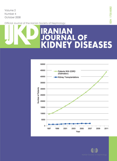
Vol. 2 No. 4 (2008)About the cover:Trend and estimation of the number of patients with end-stage renal disease (ESRD) in Iran from 1997 to 2011 (with permission from the Transplantation Management Center of Ministry of Health, unpublished data).
-
July
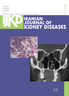
Vol. 2 No. 3 (2008)About the cover:Coronary computed tomography scan with opacification of the left-sided sinuses (maxillary, ethmoidal, and sphenoidal)-The arrow shows aseptate broad fungal hyphae within the necrotic area (hematoxylin-eosin, × 40).