
Vol. 14 No. 4 (2020): July
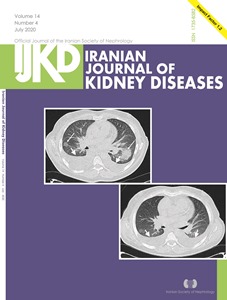
About the cover:A) A 55-year-old male patient with a history of underlying chronic kidney disease (GFR = 9.1, stage V) presented with dry cough and dyspnea which had started since 5 days ago. Initial computed tomography (CT) imaging showed bilateral moderate pleural effusion predominantly in right side, ground glass opacity and area of consolidation in middle zones. B) CT imaging obtained 22 days later show bilateral mild pleural effusion and complete lesion absorption. The patient was discharged after 7 days.
Published:
2020-07-13